Case 23Amaurosis Fugax / Carotid Emboli
Clinical Presentation
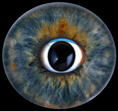
A 65-year-old man has had several episodes of momentary blindness in one eye over the past year. These episodes usually have lasted from seconds to 10 minutes. The photograph shows fundus detail of the involved eye.
Learner Questions
- Describe the ophthalmoscopic findings.
- What are the pertinent questions to be considered in the review of systems?
- What is your diagnosis?
- How would you confirm your diagnosis?
Answer Framework
Findings Diagnosis
HOLLENHORST PLAQUE - bright, glistening, yellowish-orange refractile crystalline deposit AMAUROSIS FUGAX (transient monocular vision loss - TMVL) secondary to carotid artery (cholesterol embolus) at a retinal arterial bifurcation. Appears larger than the vessel lumen embolic disease, confirmed by Hollenhorst plaque. = A RETINAL TIA. Classic: 'shade coming it occupies. Originates from carotid atherosclerotic plaque. Does not obstruct flow down over the eye' - transient, painless, monocular, seconds to minutes, complete permanently (vision returns after each episode). Retina otherwise intact. recovery. DDx: carotid emboli (this case), cardiac emboli (AF), GCA, papilledema-related TVOs (bilateral, postural), ocular migraine (young patients, positive phenomena).
ROS Confirmation
Character of VL ('shade coming down' = classic curtain effect of amaurosis fugax), Carotid duplex US (first-line - ipsilateral ICA stenosis + plaque). CTA or MRA carotid/circle duration/frequency/any permanent VL. Contralateral neurologic TIA symptoms (arm/leg of Willis (for surgical planning). TTE/TEE (cardiac source - valvular disease, intracardiac weakness, facial droop, speech difficulty). HTN, DM, hyperlipidemia, smoking, CAD, AF or thrombus). ECG + Holter monitoring (paroxysmal AF). ESR/CRP (rule out GCA in >50 years). valvular disease. Jaw claudication/scalp tenderness (GCA in elderly). Prior stroke or TIA. Fasting lipids, glucose, HbA1c, CBC.
Teaching Pearl
Amaurosis fugax = RETINAL TIA - same embolic pathophysiology as cerebral TIA, same urgency. 'Shade coming down' is classic and nearly pathognomonic. A Hollenhorst plaque on fundoscopy confirms carotid embolic disease. Management: urgent carotid imaging; CEA for symptomatic ipsilateral stenosis >=70% (NASCET trial); antiplatelet + statin + risk factor modification for all. TREAT LIKE A STROKE - same-day or next-day vascular evaluation.
Original answer transcript
Findings Diagnosis
HOLLENHORST PLAQUE - bright, glistening, yellowish-orange refractile crystalline deposit AMAUROSIS FUGAX (transient monocular vision loss - TMVL) secondary to carotid artery
(cholesterol embolus) at a retinal arterial bifurcation. Appears larger than the vessel lumen embolic disease, confirmed by Hollenhorst plaque. = A RETINAL TIA. Classic: 'shade coming
it occupies. Originates from carotid atherosclerotic plaque. Does not obstruct flow down over the eye' - transient, painless, monocular, seconds to minutes, complete
permanently (vision returns after each episode). Retina otherwise intact. recovery. DDx: carotid emboli (this case), cardiac emboli (AF), GCA, papilledema-related
TVOs (bilateral, postural), ocular migraine (young patients, positive phenomena).
ROS Confirmation
Character of VL ('shade coming down' = classic curtain effect of amaurosis fugax), Carotid duplex US (first-line - ipsilateral ICA stenosis + plaque). CTA or MRA carotid/circle
duration/frequency/any permanent VL. Contralateral neurologic TIA symptoms (arm/leg of Willis (for surgical planning). TTE/TEE (cardiac source - valvular disease, intracardiac
weakness, facial droop, speech difficulty). HTN, DM, hyperlipidemia, smoking, CAD, AF or thrombus). ECG + Holter monitoring (paroxysmal AF). ESR/CRP (rule out GCA in >50 years).
valvular disease. Jaw claudication/scalp tenderness (GCA in elderly). Prior stroke or TIA. Fasting lipids, glucose, HbA1c, CBC.
TEACHING PEARL
Amaurosis fugax = RETINAL TIA - same embolic pathophysiology as cerebral TIA, same urgency. 'Shade coming down' is classic and nearly pathognomonic. A Hollenhorst
plaque on fundoscopy confirms carotid embolic disease. Management: urgent carotid imaging; CEA for symptomatic ipsilateral stenosis >=70% (NASCET trial); antiplatelet +
statin + risk factor modification for all. TREAT LIKE A STROKE - same-day or next-day vascular evaluation.